The aim of EchoBrain (closed on 10/31/2020) is to innovate in transcranial ultrasonic imaging as an alternative to MRI or CT scans in the diagnosis of brain pathologies such as strokes, by associating two complementary partners : the Methodological Developments and Instrumentation team of Jean-Luc Gennisson, coordinator, (BioMaps) and the engineers of CEA-List who have been developing for several years an expertise platform of "Non Destructive Testing", named CIVA, which combines numerical simulation with state-of-the-art ultrasonic imaging tools.
The performance of a routine transcranial echography is very limited because the skull greatly disrupts the propagation of the ultrasonic beam, resulting in a significant degradation of the quality of the echographic image. Based on the models they have developed to simulate the propagation of ultrasonic waves in complex and solid media for non-destructive testing applications, CEA-List engineers are able to simulate the disturbances caused by the presence of the skull between the probe and the tissue to be observed. They were able to refocus the ultrasounds on the cranial soft tissues and significantly improve the quality of the echographic image.
From a methodological point of view, the adaptation of the acoustic propagation simulation tools developed by the List (CIVA) has enabled the JL Gennisson team to work on the improvement of ultrafast echographic systems. Thus, these programmable systems (photo) can take into account the acoustic aberrations of the skull simulated and calculated by CIVA. This principle of programmable ultrafast ultrasonic imaging (up to 20,000 images/s) allows the development of new ultrasonic modalities, such as elastography or ultrafast Doppler, which have already shown their interest in clinical applications, for transcranial applications.
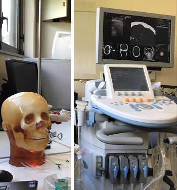
Phantom head and the programmable ultrafast US scanner (AixplorerTM, Supersonic Imagine, France) © JL Gennisson /CEA /UPSay
With EchoBrain, the researchers carried out a proof of concept for transcranial echography and showed that it was possible to partially correct aberrations due to the presence of the skull and improve the echographic image. These results, although still in their early stages, are promising for the development of ultrasonic imaging of the brain.
Joliot researcher contact : Jean-Luc Gennisson
EchoBrain website : https://attract-eu.com/showroom/project/echobrain/
Read the final project report.