The evolution of Alzheimer's disease is highly variable from one patient to another and physicians do not yet have reliable prognostic markers to predict cognitive and functional decline.
This neurodegenerative disease is characterized by the abnormal accumulation in the brain of deposits of amyloid protein on the outside of cells and of abnormal intraneuronal Tau protein constituting the neurofibrillary degenerations that modify the functioning of neurons. These protein abnormalities appear years before the onset of the first symptoms of the disease. Thanks to PET imaging and the use of the [18F]-flortaucipir tracer, it is possible to detect in vivo Tau lesions and their distribution in the brain. The topographical information provided by PET-tau imaging leads to consider this imaging method as an effective potential predictor of neurodegeneration and subsequent cognitive decline.
PREFERENTIAL ASSOCIATION BETWEEN INITIAL LOCAL TAU PROTEIN LOAD AND DECLINE IN A SPECIFIC COGNITIVE DOMAIN
Teams from GHU-Paris and the SHFJ followed 36 patients, all at an early stage of Alzheimer's disease, for two years. The researchers show that PET imaging of tau protein is predictive of the evolution of cortical atrophy, which is a consequence of Alzheimer's disease, and especially of cognitive decline. In particular, they show that decline in a specific cognitive domain is preferentially associated with initial tau tracer binding in brain regions known to be classically associated with it: for example, decline in verbal memory correlates with initial tau protein loading in the left temporal cortex.
To assess the "specificity" of tau imaging as a predictive marker of cognitive decline and atrophy progression, the researchers analyzed the predictive potential of other disease markers. Neither the initial profile of cortical atrophy on MRI, nor protein biomarkers in cerebrospinal fluid or amyloid protein imaging on PET had significant predictive value.
These results, published in the Journal of Neurology, Neurosurgery and Psychiatry, highlight the importance of tau protein in the mechanism of Alzheimer's disease and its predictive value for the progression of the disease. The use of PET imaging of tau protein could allow medical teams to better anticipate the potential evolution of cognitive disorders and to improve the management and support of patients. It could also help identify patients at high risk of progression in order to improve the design of therapeutic trials.
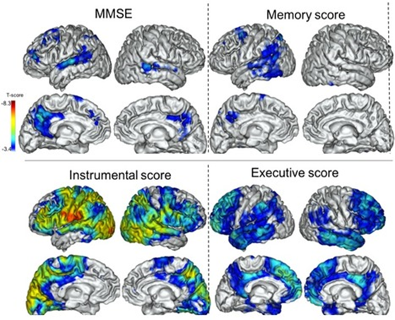
Representation of the cortical regions in which tau tracer binding is associated with the evolution of cognitive decline expressed in terms of global cognitive efficiency (MMSE), verbal memory (memory score), instrumental functions (instrumental score) and executive functions (executive score). © J. Largarde/GHU Paris
Contact GHU Paris: